
Figure 2.
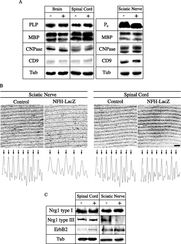
Composition and ultrastructure of myelin in NFH-LacZ mice. A, Western blot analysis of myelin associated proteins. The crude myelin extracts from brain, spinal cord, and sciatic nerve of 3-month-old control (−) and NFH-LacZ (+) mice were analyzed by Western blot using antibodies recognizing P0, PLP, MBP, CNPase, CD9, and tubulin (Tub) for control. The two bands shown for MBP are the 14 and 17 kDa isoforms and were combined for quantification. The amount of these proteins was unaffected in transgenic mice, except for P0 that was decreased by 23 ± 8% in NFH-LacZ sciatic nerves. B, Electron micrographs showing periodicity of myelin in the sciatic nerve and the spinal cord of 3-month-old mice. Scale bar, 20 nm. Densitometry graphs described the myelin period in sciatic nerve and spinal cord from control and transgenic mice. Arrows indicate deflections corresponding to major dense lines. Note the difference of lamellar spacing between control and NFH-LacZ sciatic nerves. C, Western blot analysis of Nrg1 types I and III, and erbB2 in total protein lysates from control and NFH-LacZ sciatic nerve and spinal cord. Expression of Nrg1 type III is reduced in NFH-LacZ sciatic nerve whereas erbB2 is increased in NFH-LacZ spinal cord.