
Figure 4.
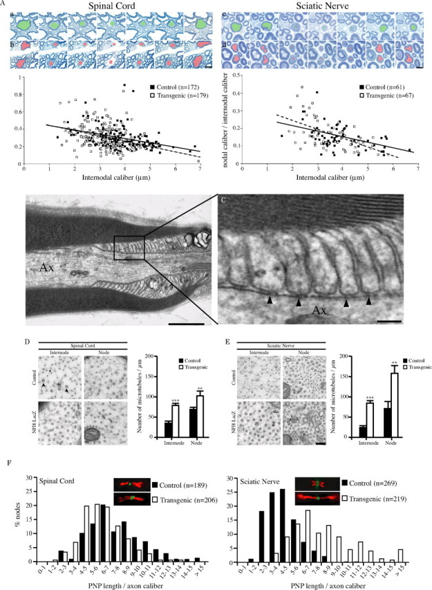
Despite the absence of NFs in internodal segments, the axonal caliber is reduced at nodes of Ranvier. A, Serial sections of spinal cord (a, b) and sciatic nerve (c, d) from 3-month-old control (a, c) and NFH-LacZ mice (b, d). Despite the absence of axonal NF in internodal segments of transgenic samples, the extent of the axonal caliber reduction at the nodes of Ranvier is similar between transgenic and control fibers. Bottom, The level of axonal caliber reduction at nodes is more important for large fibers than for small fibers, both in control (black squares) and NFH-LacZ (white squares) samples, and in the PNS as well as in CNS. Regression lines are represented (full, control; dotted, NFH-LacZ). B, C, Electron micrographs through the paranodal region (longitudinal section) from a 4-month-old NFH-LacZ mouse L4 ventral root. Axon (Ax) caliber is reduced despite the absence of detectable NFs. Note the presence of typical transverse bands (arrowheads) indicating the correct formation of the junctional complex normally formed between the axolemma and paranodal loops. D, E, Axons with NFs or deprived of NFs have an increased density of microtubules in both nodal and internodal domains, as observed on ultrathin sections of 3-month-old control and NFH-LacZ spinal cords (D) and sciatic nerves (E). Arrowheads point toward the MT whereas the asterisk indicates NF. Error bars indicate SEM. **p < 0.01; ***p < 0.0001. F, Ratio between PNP length and axon caliber was evaluated in sciatic nerve and spinal cord from control and NFH-LacZ mice using anti-Caspr labeling. This ratio is increased in transgenic sciatic nerves compared with control samples, suggesting that the length of paranodes is inappropriately long for these smaller axons. However, the ratio is slightly reduced in transgenic spinal cord. Scale bars: A, 2 μm; B, 1 μm; C, 0.1 μm; (in E) D, E, 100 nm.