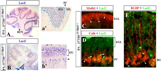
Figure 1.
IFT88 expression in the adult and developing mouse cerebellum. Direct lacZ (A, B) and antibody staining (C–E) of sagittal cerebellar sections of IFT88 Δ2–3βgal/+ mice at the indicated stages. Insets (a′, b′) show higher magnification of boxed regions. Deep cerebellar nuclei (DCN), Purkinje cells (PC), molecular layer (ML), internal granule cell layer (IGL), external granule cell layer (EGL), and Bergmann glia (BG) are indicated. b′, Black arrowhead points to the location of Purkinje cells and Bergmann glia. C–E, White arrowheads point to LacZ+ dots in Math1+ granule cells (C), calbindin+ Purkinje cells (D), and BLBP+ Bergmann glia (E).

