
Figure 6.
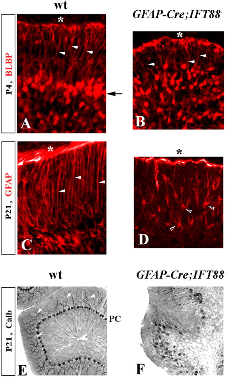
Bergmann glia and Purkinje cell defects in hGFAP–Cre+;IFT88floxed/null mutant mice. Immunostained sections of wild-type (wt) (A, C, E) and hGFAP–Cre+;IFT88floxed/null mutant (B, D, F) cerebella at the indicated stages. A–D, Arrow points to Bergmann glia aligned along Purkinje cells in the wild-type but not in the hGFAP–Cre+;IFT88floxed/null cerebellum. White arrowheads point to radial glia fibers extending to the cerebellar pial surface (asterisk). Black arrowheads point to mutant Bergmann glia with astrocyte-like morphology. E, F, Arrowheads point to wild-type Purkinje cell (PC) dendrites, which are not recognizable in hGFAP–Cre+;IFT88floxed/null cerebellum. calb, Calbindin.