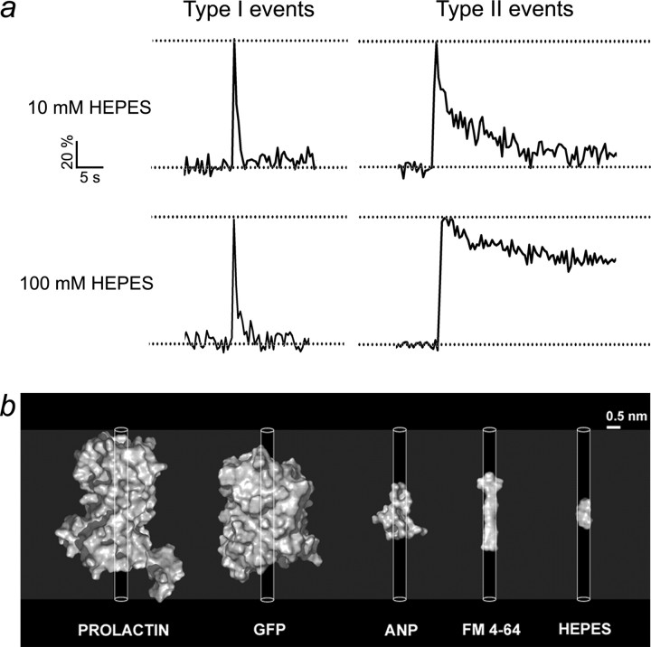
Figure 4.
Addition of small HEPES molecules to the bath slows reacidification only in vesicles that load FM 4-64. a, Representative spontaneous type I and type II exocytotic events in 10 and 100 mm HEPES bath solution. spH fluorescence intensity was normalized as (F − F0)/(Fmax − F0), where F is the fluorescence at any given time point. Dotted lines indicate minimal and maximal levels of spH fluorescence signal. Note the slower spH fluorescence decline of type II events in the 100 mm HEPES bath solution. b, Surface models of prolactin, green fluorescent protein (GFP), atrial natriuretic peptide (ANP), FM 4-64, and HEPES. The three-dimensional (3D) structure data for prolactin, GFP, and ANP were from Brookhaven Protein Data Bank (PDB). The 3D structure data for HEPES were from the National Cancer Institute database. The 3D structure of FM 4-64 was predicted from the two-dimensional structure using MDL ISIS/Draw (version 2.5). The surface models of molecules were generated with a Pymol Molecular Graphics System. The cylinders indicate the dimension of a fusion pore with a diameter of 0.5 nm and a length equal to the membrane thickness. See Table 3 for dimension.

