
Figure 1.
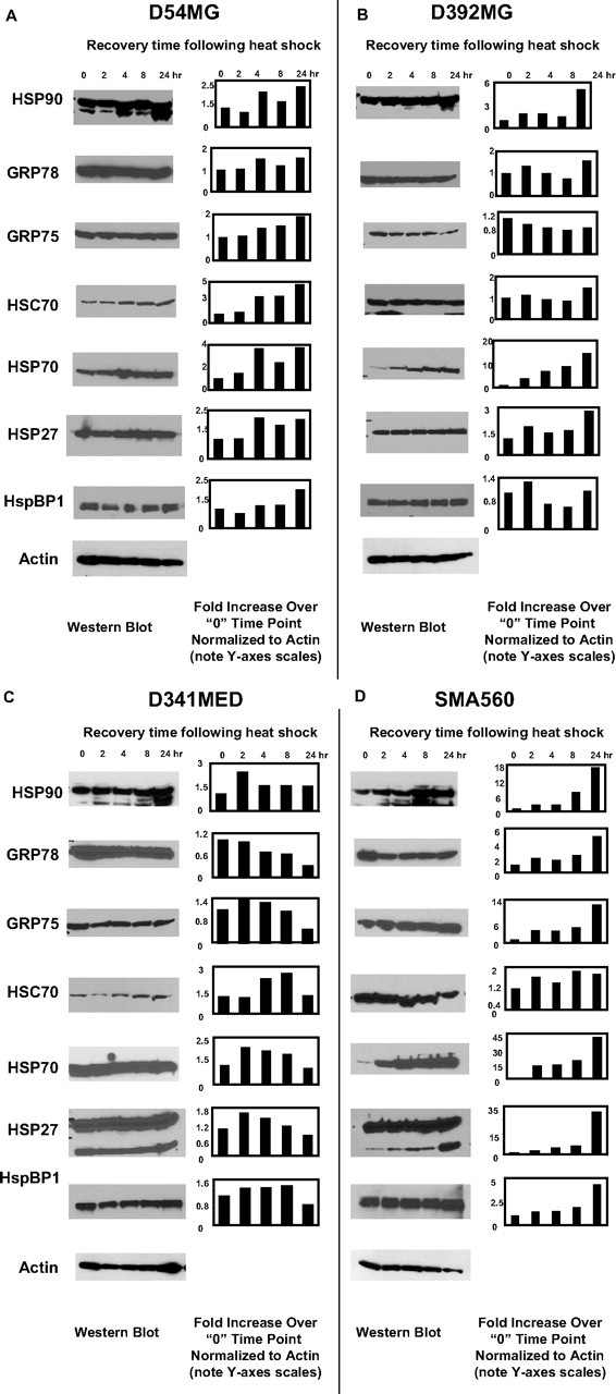
Heat shock generally increases chaperone/heat shock protein expression in brain tumor cell lines. A–D, Cell lines derived from patient GBMs (D54MG, A; D392MG, B), from a medulloblastoma (D341MED, C), or from a murine astrocytoma (SMA560, D) were subjected to sublethal heat shock (42°C, 1 h) and allowed to recover for the time periods indicated after heat shock (2–24 h). Lysates were prepared from cells at the time points indicated (“0” refers to before heat shock); proteins were separated by LDS-PAGE and Western blotted. Antibody probes are listed on the left. Quantitative comparisons (bar graphs) were made by densitometry relative to actin staining (average of two independent experiments). Error bars indicate SD. All other SDs were <10%. Cells were >95% viable whether heat shocked or not.