
Figure 1.
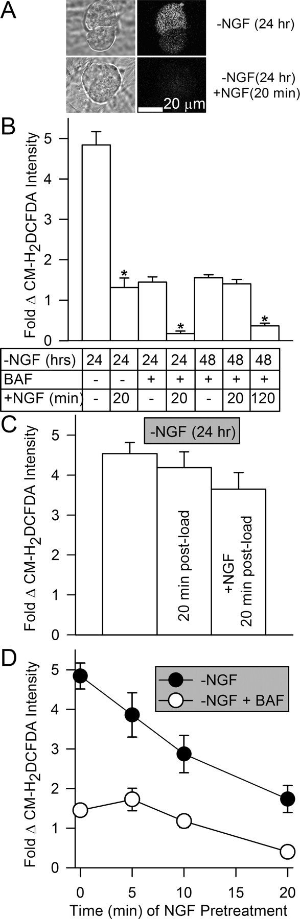
Elevated levels of ROS in NGF-deprived mouse sympathetic neurons in cell culture were rapidly suppressed by re-exposure to NGF. A, Paired phase and confocal micrographs of neurons loaded with the redox-sensitive dye CM-H2DCFDA. By 24 h after NGF withdrawal, CM-H2DCFDA intensity had increased, indicating that the dye was oxidized by ROS. Reintroduction of NGF during the 20 min period of CM-H2DCFDA loading inhibited this oxidation. B, Effects on neuronal CM-H2DCFDA intensity of NGF readdition to cultures deprived of NGF for 24–48 h. Because of ongoing apoptotic death, all of the cells deprived of NGF for the longer period were maintained alive by adding the broad-spectrum caspase inhibitor BAF (50 μm) to the culture medium at the time of NGF withdrawal. Note that the caspase inhibitor also potently suppressed ROS (e.g., CM-H2DCFDA intensity) (Kirkland et al., 2002a,b). For neurons deprived of NGF for 24 h, NGF readdition was done during the 20 min period of CM-H2DCFDA loading. The 48 h BAF-supported neurons were re-exposed to NGF either during the 20 min dye-loading period or for 100 min before dye loading plus the 20 min of dye loading. Stars indicate a significant difference (p < 0.001 by ANOVA) from the same condition with no readdition of NGF during the period of CM-H2DCFDA loading. n = 62–588 neurons. C, NGF readdition did not suppress CM-H2DCFDA intensity in NGF-deprived neurons by reducing the dye after it had become oxidized. Cultures deprived of NGF for 24 h were loaded with CM-H2DCFDA for 20 min. The first bar is control CM-H2DCFDA intensity at the end of the 20 min load. For the other two conditions, the dye was washed out after the 20 min load either with medium containing NGF or lacking NGF. Dye intensity was then determined 20 min later. n = 126–522 neurons. D, Time course of recovery of elevated ROS after NGF readdition followed by subsequent withdrawal. ROS recovered more slowly when NGF was again withdrawn after longer periods of NGF readdition. Neurons deprived of NGF for 24 h (±50 μm BAF) were exposed to NGF for various times before being loaded with CM-H2DCFDA for 20 min in medium containing no NGF and a NGF-neutralizing antibody. Fold change shown in this and subsequent figures is change from the intensity of the dye measured in sibling cultures of neurons maintained since the time of plating in NGF-containing medium. n = 104–350 neurons.