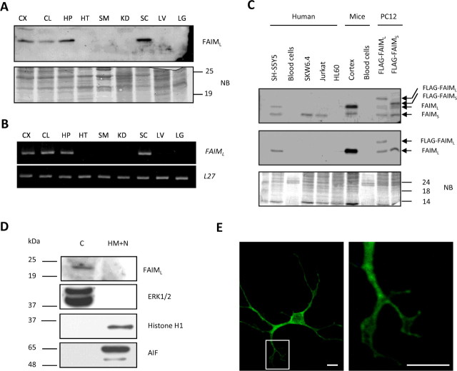
Figure 1.
FAIML is expressed only in the nervous system. A, Adult mouse tissue lysates (20 μg) were analyzed by SDS-PAGE/immunoblot using anti-FAIML serum (1:1000) (top panel). As the loading control, the membrane was stained with Naphtol Blue. B, Mouse FAIML mRNA tissular content was assessed by semiquantitative RT-PCR with specific primers and compared with L27 housekeeping gene. C, Immunoblot analysis of the FAIML expression in human and mouse hematopoietic/immune cells as indicated. Cortical neurons, neuroblastoma SH-SY5Y, and transfected PC12 extracts were used as a positive control of the FAIML and total FAIM expression. Twenty micrograms of total protein were resolved by SDS-PAGE and blotted with anti-FAIMS (top panel) and FAIML (middle panel). Naphtol blue staining was used as a loading control (bottom panel). D, PC12 cells were lysed in a hypotonic solution, and two crude subcellular fractions were prepared. The equivalent protein content of each fraction was subjected to SDS/PAGE immunoblot analysis using antibodies specific for FAIML, pan-ERK (cytosolic marker), Histone H1 (nuclear marker), and AIF (mitochondria marker). C, Cytosolic fraction; HM+N, heavy membrane and nuclear fraction. E, Subcellular distribution of FAIML. Confocal microscopy (60×) of E15 cortical cells transfected with FLAG-FAIML and immunofluorescence with anti-FLAG coupled to a secondary anti-mouse conjugated to FITC. Scale bar, 20 μm. CX, Cortex; CL, cerebellum; HP, hippocampus; HT, heart; SM, skeletal muscle; KD, kidney; SC, spinal cord; LV, liver; LG, lung; NB, Naphtol Blue; C, cytosolic; HM, heavy membranes; N, nuclei.

