
Figure 8.
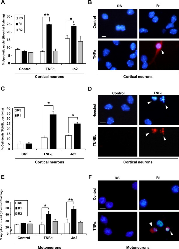
Endogenous FAIML, but not FAIMS, is responsible for the resistance of primary neurons to DR activation. A, E15 cortical neurons were infected with R1, R2, or RS for 72 h before Fas-Jo2 (5 μg/ml) and TNFα (100 ng/ml) treatment. Twenty-four hours later, apoptotic cell death was determined by scoring the percentage of apoptotic cells after Hoechst staining. B, E15 cortical neurons were treated as in A. Twenty-four hours later, active caspase-3 was detected by immunofluorescence. Images show active caspase-3 immunofluorescence (red) merged with Hoechst staining. C, E15 cortical neurons were infected and treated as indicated in A. Then, apoptosis was assessed by the TUNEL assay. D, Representative images of C. E, E12.5 mice motoneurons were infected with lentiviruses for 5 d. Then, cells were treated with Fas-Jo2 (1 μg/ml) or TNFα (100 ng/ml) for an additional 24 h. Percentage of cell death was measured by Hoechst staining of nuclei and counting apoptotic profiles. F, Immunofluorescence of active caspase-3. Images are the result of merging Hoechst staining with active caspase-3 immunofluorescence. Ctrl, Control. *p ≤ 0.05; **p ≤ 0.01. Scale bars, 25 μm. Arrowheads indicate apoptotic cells (B, F) and nuclei (D).