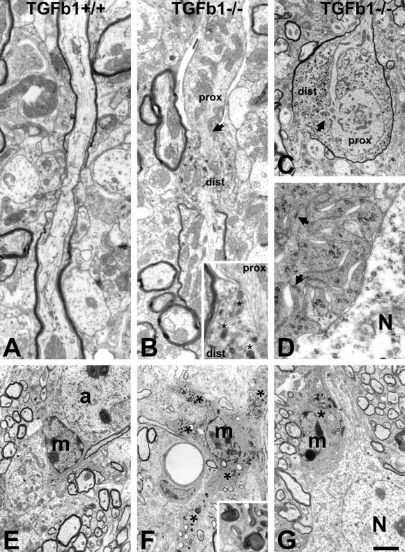
Figure 3.

Ultrastructural changes in the brain of TGFβ1-deficient, −/− mice. A, E, Normal axons, microglia (m), and astrocytes (a) in the control, TGFβ1+/+ animals. B, C, TGFβ1−/− mice show frequent disturbances in axonal transport. These disturbances lead to the local bulging of axons to accommodate the anterogradely and retrogradely transported material, particularly mitochondria, in the proximal (prox) and distal (dist) parts of the axon, respectively. Affected axons sometimes become partly demyelinated (proximal segment). As shown in B (inset), microtubular structure at the site of interruption (arrows) is frequently disorganized and displaced. Retrogradely transported material accumulating in the distal part also contains large and round breakdown organelles (asterisks) that may represent mitochondrial fragments. D, F, G, The microglia in TGFβ1−/− mice consistently show areas with numerous, moderate-size phagocytic inclusions (asterisks and F, inset) that extend into swollen proximal processes (F). They also exhibit an abnormal attachment to neighboring neurons (N) and, at higher magnification, many membranous ruffles with frequent, homophilic focal adhesions (D). Scale bar: (in G) A, B, 0.8 μm; B inset, 0.3 μm; C, 1.5 μm; D, 0.4 μm; E–G, 2.5 μm.
