Figure 4.

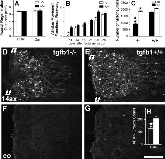
TGFβ1 and axonal regeneration, sprouting, functional recovery, and neuronal cell death after facial axotomy. A, No effect on the regeneration distance of CGRP- and galanin (Galn)-immunoreactive motor axon populations, 4 d after facial nerve crush (mean ± SEM; n = 6 animals per group). B, Functional recovery of whisker hair movement after facial nerve cut (n = 10 +/+ and 7 −/− animals). *p < 0.05 in unpaired Student's t test (uSTT) between +/+ and −/− groups. C, Changes in neuronal cell number in the axotomized (ax) and contralateral (co) facial motor nucleus, 30 d after a left facial nerve cut. Absence of TGFβ1 enhances neuronal cell disappearance after axotomy, from 13% of the injured neurons in the +/+ to >52% in the −/− animals. Note the >10% smaller neuronal cell number on the unoperated, contralateral side in the TGFβ1-deficient mice. n = 9 +/+ and 6 −/− mice; *p < 0.025 in uSTT between the same side in +/+ and −/− mice; #p < 0.001, uSTT for the difference in the ratio of operated/unoperated side in +/+ to −/− animals. D–H, Central sprouting of galanin-immunoreactive axons in TGFβ1−/− (D, F) and +/+ facial nuclei (E, G), 14 d after facial nerve cut (14ax); co, contralateral side (C, D); H shows the quantification of the growth cones per tissue section (n = 3 animals per group). Under normal conditions, facial axotomy causes an induction of neuronal galanin immunoreactivity and a proliferation of galanin-immunoreactive axonal sprouts, particularly in the white matter marked by double arrows just ventral to the facial motor nucleus, peaking 14 d after facial nerve cut. This axonal sprouting is strongly curtailed in the TGFβ1−/− mice (*p < 0.05, uSTT). AFMN, Axotomized facial motor nucleus. Scale bar, 250 μm.
