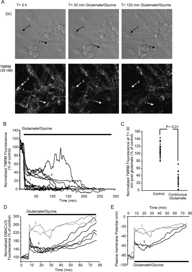
Figure 4.
Prolonged glutamate excitation induces a rapid loss of ΔΨm and early necrotic injury in cerebellar granule neurons. Cerebellar granule neurons plated on Willco dishes were loaded with 30 nm TMRM and continuously exposed to glutamate/glycine (100 μm/10 μm). TMRM fluorescence was then monitored over time, and images were taken at 1 min intervals. A, Differential interference contrast (DIC) and TMRM fluorescent images were chosen at selected time points (0, 30, and 120 min during glutamate excitation) from a representative experiment. B, Representative traces for whole-cell TMRM fluorescence in neurons during prolonged glutamate excitation. Two major subgroups were identified: traces that show a rapid collapse of ΔΨm (i) and traces that show a partial recovery of ΔΨm, followed by a secondary collapse of ΔΨm (ii). C, Whole-cell TMRM fluorescence in control neurons after 30 min and neurons treated with glutamate for 30 min. p < 0.001, difference between TMRM fluorescence for control neurons and neurons after 30 min continuous glutamate excitation (control, n = 39; necrotic, n = 63). D, Cerebellar granule neurons were loaded with the ΔΨp-sensitive probe DiBAC2(3) (1 μm) and continuously exposed to glutamate/glycine (100 μm/10 μm). Neurons undergo a rapid monophasic (i) collapse of ΔΨp or a biphasic (ii) collapse of ΔΨp (traces are representative of those obtained from 3 separate experiments). E, Representative traces for modeled changes in ΔΨp (C) for neurons undergoing a rapid monophasic (i) collapse of ΔΨp or a biphasic (ii) collapse of ΔΨp when continuously exposed to glutamate/glycine (100 μm/10 μm).

