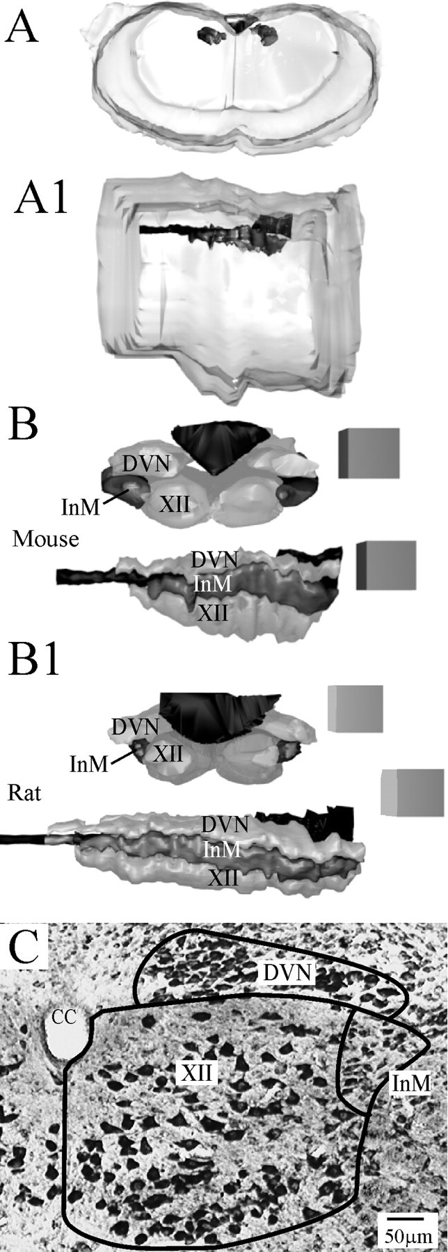
Figure 1.

Serial reconstruction of the InM. A, A1, Guide images showing an approximate outline of the surface of the mouse medulla oblongata and the position of the central canal/fourth ventricle in relation to this, as viewed from either a more rostral location (A) or a lateral view (A1). The DVN, XII, central canal, and InM are the darker areas at the dorsal aspects of the diagram. A zoom-in on this region is shown in the panels below for both mouse and rat. B, B1, 3D reconstructions from serial toluidine blue-stained mouse (B) and rat (B1) tissue viewed from a rostral position shows that the InM (dark gray) lies slightly lateral to the XII and ventral to the DVN. Looking from a lateral position, the reconstructions show that all three structures have a similar rostrocaudal extent. All edges of cubes in reconstructions are 300 μm. C, Representative micrograph of a toluidine blue section indicating the anatomical boundaries of the InM used in the reconstruction. CC, Central canal.
