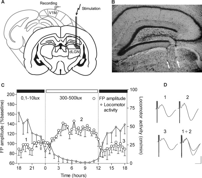
Figure 1.
Basal synaptic transmission in the primary visual cortex of freely moving animals. A, Illustration of the position of the bipolar stimulating electrode at the dLGN and monopolar recording electrode in the primary visual cortex. B, Nissl-stained coronal section showing stimulation electrode tracks that reach dLGN. White arrows point to the location of electrode tips. C, Daily oscillations of field potential amplitude in V1 during a 12 h light/dark cycle. After I/O curve measurement, baseline recording was started in conditions of dark exposure (ZT 18). The switch to daylight illuminance levels induced a suppression of the animals' locomotor activity. The locomotor activity is measured as distance in centimeters/minute. D, Analogs represent FPs evoked at the points marked here and in subsequent figures. Calibration: 5 ms, 1 mV.

