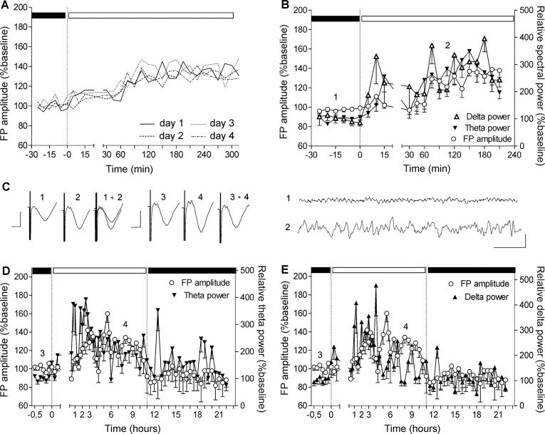
Figure 2.
Slow-frequency rhythms increase concurrently with the augmentation of the basal synaptic transmission. A, Baseline recordings from a group of animals (n = 4) conducted in different sessions. The FP augmentation has the same profile that is independent from the day of recording. For clarity of the figure, the SEs of the average means are not presented. B, EEG recorded from VC during the switch between low and high illuminance of the dark/light cycle. Elevation of the slow theta and delta frequencies is preceding the FP amplitude elevation and the transition of the animal into the inactive behavioral state. C, Left analogs represent FP traces evoked at the points marked in the figures. Calibration: 5 ms, 1 mV. Right analogs represent low-pass filtered EEG analog traces from epochs marked in the figures. Calibration: 1 s, 0.1 mV. Theta (D) and delta (E) power elevation continues during the first 3–4 h of the subsequent behaviorally inactive period. After stabilization of the augmented FP amplitude, delta and theta frequencies diminish their elevation, which reaches baseline levels during the following activity state (in close association with the return of FP amplitude to basal levels).

