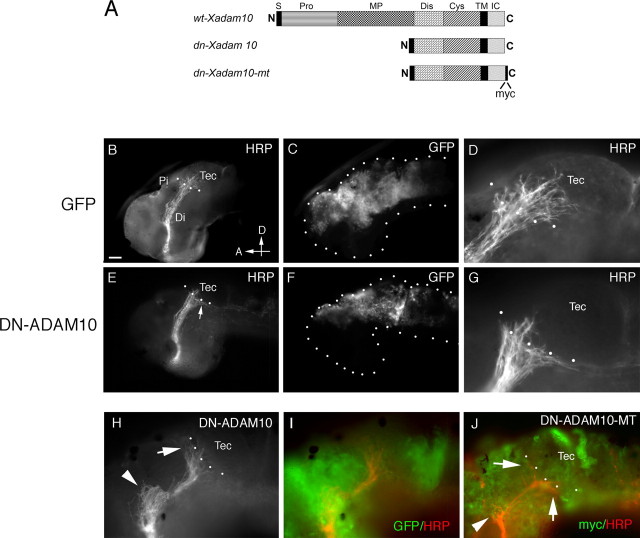
Figure 5.
Misexpression of DN-ADAM10 in the dorsal neuroepithelium causes target recognition defects. A, Schematic diagram of the DNADAM10 constructs used in the electroporation and transfection experiments. S, Signal sequence; Pro, prodomain; MP, metalloproteinase domain; Dis, disintegrin domain; Cys, cysteine-rich region; TM, transmembrane domain; IC, intracellular domain; N, N terminal; C, C terminal. B–J, Transgene-expressing cells and HRP-labeled optic projections in lateral views of stage 40 brains. mRNA for GFP (B–D), GFP+DNXadam10 (E–I), and DNXadam10-mt (J) were injected into the anterior brain vesicles and electroporated into the dorsal neuroepithelium. At stage 40, the optic projections were anterogradely labeled with HRP, and the brains were processed for whole-mount immunochemistry with anti-HRP and anti-GFP or anti-myc. D and G are high-power views of the images shown in B and E, respectively. Note that many more axons enter the optic tectum in the control (D) than in the DNADAM10-expressing (G) brain. H shows an HRP-labeled optic projection in a brain electroporated with GFP+DNXadam10, and I illustrates the same projection along with expression of the coelectroporated GFP transgene. J shows immunolabeling with an antibody against the myc tag to visualize DNXADAM10-MT expression (green) and an antibody against HRP to visualize RGC axons (red). The arrows and arrowheads in H and J indicate target recognition and turning defects, respectively. The dotted lines outline the embryonic brain (C, F) and indicate the approximate anterior border of the optic tectum (B, D, E, G, H, J). Di, Diencephalon; Pi, pineal gland; Tec, tectum; D, dorsal; A, anterior. Scale bar: B, C, E, F, 50 μm; D, G–J, 25 μm.

