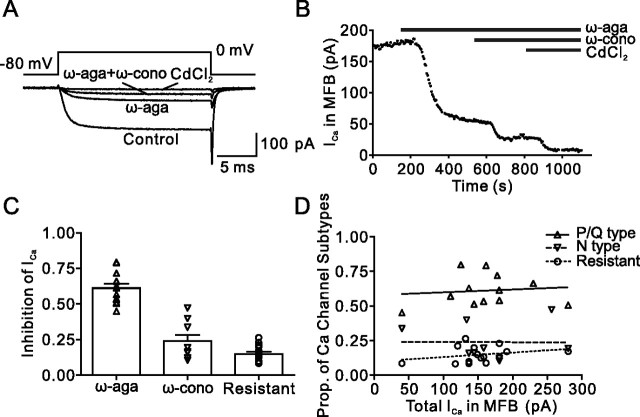
Figure 1.
Pharmacological dissection reveals three components of the presynaptic Ca2+ current in hippocampal MFBs. A, Presynaptic Ca2+ current evoked by a 20 ms pulse to 0 mV under control conditions, in the presence of 500 nm ω-agatoxin IVa (ω-aga), and after subsequent addition of 1 μm ω-conotoxin GVIa (ω-cono). The residual component was blocked by 200 μm Cd2+, confirming that it was mediated by Ca2+ channels. B, Plot of Ca2+ current amplitude against time during application of the toxins. The time of application of the blockers is indicated by the horizontal bars. Same bouton as shown in A. C, Summary bar graph illustrating the proportions of ω-agatoxin IVa-sensitive, ω-conotoxin GVIa-sensitive, and toxin-resistant component of the Ca2+ current. Symbols (triangles, inverted triangles, and circles) represent individual experiments, and bars represent mean ± SEM. Data are from 12, 9, and 15 boutons. D, Plot of the proportions (Prop.) of the three components against the total Ca2+ current amplitude. Note the lack of correlation (Pearson's r, 0.11, −0.01, and 0.28; p > 0.2 in all cases).

