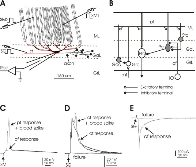
Figure 1.
Local circuitry and experimental arrangement. A, Schematic drawing of a Purkinje cell and a single climbing fiber, oriented in the parasagittal plane. The smooth, proximal dendrites of the ganglionic layer give rise to the long, spine-covered dendrites of the molecular layer. The molecular layer dendrites are primarily unbranched and traverse the molecular layer in parallel to each other. The climbing fiber (shown in red) terminates only on the soma and smooth dendrites of the ganglionic layer. SM1 and SM2, Stimulation of molecular layer to activate parallel fibers; SG, stimulation of deep layer to activate climbing fibers; Rec, recording. B, Diagram of the local circuitry of the central lobes of the mormyrid cerebellum. Some essential features of the local circuitry are shown. The climbing fiber terminates in the ganglionic layer and does not enter the molecular layer. The Purkinje cell terminates locally on the efferent cell. Parallel fibers excite the molecular layer dendrites of efferent cells, as well as the dendrites of Purkinje cells, Golgi cells, and stellate cells. Inhibitory neurons are shown in gray. cf, Climbing fiber; Efc, efferent cell; GaL, ganglionic layer; Goc, Golgi cell; Grc, granule cell; GrL, granule layer; IO, inferior olive; mf, mossy fiber; ML, molecular layer; Pc, Purkinje cell; pf, parallel fiber; Stc, stellate cell. C, Responses of a Purkinje cell to parallel fiber activation by SM in current-clamp configuration. An all-or-none broad spike arises from the parallel fiber-evoked EPSP on one sweep. D, Responses of a Purkinje cell to climbing fiber activation by SG in current-clamp configuration. The all-or-none CF-evoked EPSP can evoke a broad spike. E, Responses of a Purkinje cell to climbing fiber activation by SG in voltage-clamp configuration. The CF evokes a large all-or-none EPSC. Note that small EPSCs or EPSPs are visible in D and E when climbing fiber activation failed, perhaps caused by activation of a small number of ascending granule cell axons by SG.

