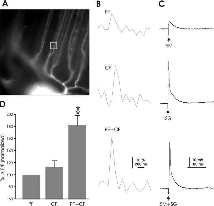
Figure 12.
Calcium transients induced by paired parallel fiber and climbing fiber activation. A, Image of the dye-filled Purkinje cell that was used for the recordings shown in B and C. The white box indicates the ROI. B, Calcium transients observed with PF stimulation (top), CF stimulation (middle), and paired PF+CF stimulation (bottom). C, Corresponding electrical traces recorded in current-clamp mode (SM, parallel fiber stimulus to molecular layer; SG, climbing fiber stimulus to ganglionic layer). D, For each recording, calcium signal amplitudes reached by PF stimulation were normalized to 100% (left bar). Calcium signal amplitudes reached with CF and paired stimulation were expressed as relative values normalized to the levels reached by PF activation. **p < 0.01.

