Figure 8.
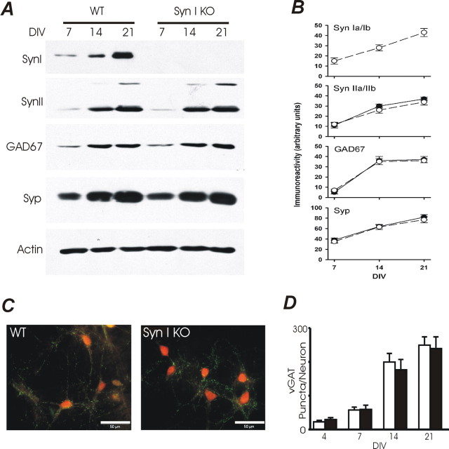
Expression of presynaptic proteins and number of GABAergic synaptic contacts are not affected by SynI deletion in hippocampal neurons during in vitro differentiation. A, Protein extracts (10 μg/lane) from WT and SynI KO hippocampal neurons were prepared at the indicated DIV and analyzed for the expression levels of synapsin Ia/Ib (SynI), synapsin IIa/IIb (SynII), GAD67, and synaptophysin (Syp). A representative immunoblot is shown. Equal loading was confirmed by the constant actin levels. B, Immunoblots from three independent replications of the experiment shown in A were quantitatively analyzed by densitometric scanning of the fluorograms. The developmental patterns of expression of SynI, SynII, GAD67 and Syp in WT (open symbols) and KO (closed symbols) neurons as a function of the days in vitro (DIV) are plotted as means ± SE in arbitrary units. C, Hippocampal neurons from WT (left panel) or Syn I KO (right panel) mice were fixed at 21 DIV and immunostained for V-GAT (green) and NeuN (red). V-GAT-positive puncta were counted and normalized by the cell number. Scale bar: 50 μm. D, The total number of inhibitory terminals in WT (white bars) and SynI KO (black bars) cultures was obtained by automatic counting and divided by the number of neurons in the field (10–15 images for each age in culture and from at least 3 preparations conducted in parallel). No significant differences in the density of GABAergic terminals were observed between WT and KO neurons at the various DIV analyzed.

