Figure 2.
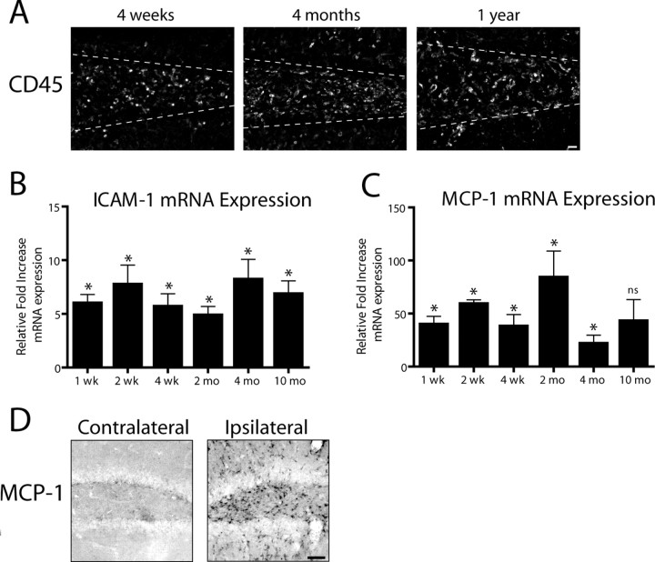
Induction of IL-1β expression drives chronic leukocyte recruitment to the mouse hippocampus. IL-1βXAT line B/b mice received unilateral intrahippocampal injections of FIV-Cre and were analyzed over a prolonged time course. A, CD45 staining in the ipsilateral (injected) hemispheres at the times indicated after FIV-Cre injection identifies infiltrating leukocytes. A white dashed line approximates the location of the granule cell layer of the dentate gyrus. qRT-PCR (B, C) generated a ratio of gene expression in the ipsilateral hippocampi of FIV-Cre injected IL-1βXAT mice compared with wild-type control animals at the same time point for ICAM-1 (B) and MCP-1 (CCL2) (C). D, Immunohistochemical detection of MCP-1 protein expression in the ipsilateral and contralateral hemispheres 2 weeks after FIV-Cre. n = 3–5 animals per time point. Scale bars: A, 25 μm; D, 50 μm. Graphs represent mean ± SEM. ns, Not significant. *p ≤ 0.05.

