Figure 3.

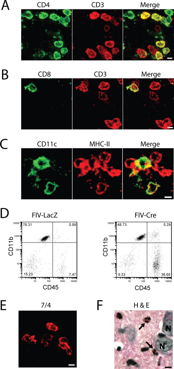
IL-1β expression recruits diverse leukocyte populations to the mouse hippocampus. Leukocyte populations were defined in the ipsilateral hippocampi of IL-1βXAT line B/b mice 2 weeks after gene activation by immunohistochemistry (A–C, E), flow cytometry (D), and tissue staining (F). A, B, T-cell populations were identified and classified by their expression of CD3 (red) and either CD4 (A; green) or CD8 (B; green), where coexpressing cells appear yellow in merged images. C, The presence of dendritic cells was established by expression of both CD11c (green) and MHC-II (red), appearing yellow in merged images. D, An increase in the number of infiltrating macrophages is demonstrated 2 weeks after FIV-Cre versus FIV-LacZ injection by expansion of the CD45hi, CD11b+ cell population. E, F, Neutrophils were identified by cellular labeling with the 7/4 antibody (E) and after hematoxylin and eosin (H&E) tissue staining (F, arrows). N, Neuronal nuclei. Scale bars, 5 μm.
