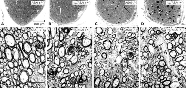
Figure 8.

Analysis of the optic nerve. Light micrographs (A–D; semithin sections, toluidine blue) and electron micrographs (E–H). Genotypes as indicated (transgenic line tg2645). It has to be mentioned that, except for ASA(+/−), the retinas of all respective optic nerves shown here displayed degeneration of the photoreceptor cells. The optic nerve of ASA(+/−) mice (A) was inconspicuous; the nerve fibers appear densely packed (E). The optic nerve in tg/ASA(+/−) (B) is inconspicuous at the LM level; at the EM level astroglial processes (A) appear slightly increased. The non-tg ASA(−/−) nerve (C) contains several sulfolipid-storing macrophages (arrows) such as shown in Figure 4, D and E. At the EM level, astrogliosis is apparent in ASA(−/−) mice (G). The arrow points to a thinly myelinated axon of relatively large diameter, suggestive of remyelination. In the tg/ASA(−/−) nerve (D), the sulfolipid-storing macrophages are even more increased in size and number. At the EM level, astrogliosis (A) is apparent (H); arrows point to thinly myelinated axons, and the double arrow points to an axon without myelin sheath despite its relatively large diameter, suggestive of remyelination and demyelination, respectively. Scale bars: A–D, 100 μm; E–H, and 2.5 μm.
