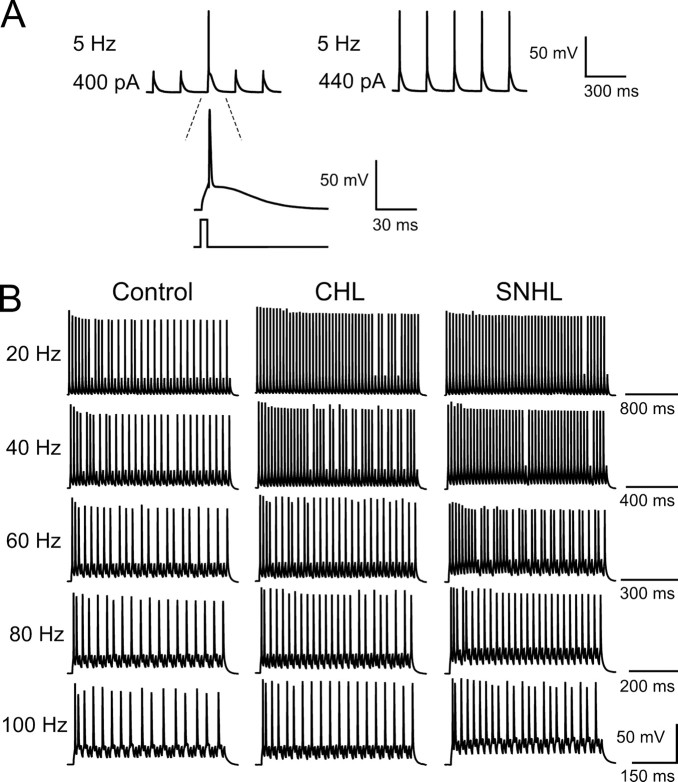
Figure 5.
Hearing loss decreases frequency-dependent spike adaptation. A, For all neurons, pulse amplitude was set to produce 100% spike response (top trace). As pulse amplitude increased, evoked action potentials became more reliable. A voltage response from a control neuron to a 5 Hz train of current pulses delivered at 400 or 440 pA is shown. B, Hearing-loss neurons show less spike adaptation. Left, Discharge pattern of a control neuron in response to train current injection of 50 pluses at 20–100 Hz. The spike numbers are 30, 28, 22, 19, and 16 for 20, 40, 60, 80, and 100 Hz, respectively. The resting membrane potential is −64 mV. Middle, Discharge pattern of a CHL neuron in response to train current injection of 50 pluses at 20–100 Hz. The spike numbers are 47, 40, 28, 24, and 19 for 20, 40, 60, 80, and 100 Hz, respectively. The resting membrane potential is −63 mV. Right, Discharge pattern of a SNHL neuron in response to train current of 50 pluses at 20–100 Hz. The spike numbers are 48, 47, 37, 27, and 22 for 20, 40, 60, 80, and 100 Hz, respectively. The resting membrane potential is −65 mV.

