Figure 6.
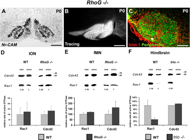
RhoG mutant mice show a normal organization and projections of IONn and a proper development of fMN. Quantification of Rho GTPases activity in E15 wild-type mice and RhoG and trio mutant mice. Nr-CAM in situ hybridization revealed a normal organization of the rostral ION in RhoG mutant mice at P0 (A). Retrograde tracing of ION allowed the visualization of the whole contralateral IO complex (B). The immunostaining with anti-Peripherin (in green) and anti-Islet-1 (in red) revealed the distinct fMN subnuclei (C). Quantification of the relative active state of Rac1 and Cdc42 after Western blot quantification in both ION (D) and fMN (E) extracts. The rate of active Rac1 and Cdc42 did not change significantly in RhoG mutant mice (p > 0.1) in either ION extracts (D) or fMN extracts (E). Western blot analysis after GST pull down was also performed on individual entire hindbrain for WT and trio mutant mice (F). The activity of Cdc42 remained unchanged in trio mutant mice, but the activity of Rac1 showed a 80% decrease. Scale bars: A, 120 μm; B, 140 μm; C, 180 μm.

