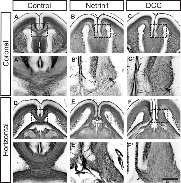
Figure 1.

Probst bundles in E18 DCC and Netrin1 mutant brains. Immunostaining of coronal and horizontal sections from E18 Netrin1 and DCC mutant brains and wild-type controls with GAP43 antibody, an axon marker, and visualization by a nickel–DAB chromagen are shown. A, D, Control sections show normal formation of the corpus callosum in the coronal (A) or horizontal (D) view. B, C, E, F, In contrast, Probst bundles can be clearly observed in sections from either Netrin1-deficient (B, E) or DCC-deficient (C, F) brains (boxes in A–F indicate the areas shown in higher power in A′–F′). After immunohistochemistry, where the entire Probst bundle is stained, swirling fibers can be observed within the bundle, especially in the coronal view (B′, C′, arrows) and in the ventral region of the bundle. Scale bar: (in F′) A–F, 1 mm; A′–F′, 250 μm.
