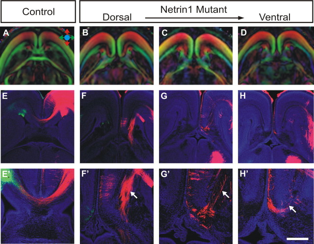
Figure 3.
DTMRI and tract-tracing analysis of Probst bundles in Netrin1 mutants. Using both DTMRI scanning and carbocyanine dye tract tracing, the fiber structure of Probst bundles were analyzed in E18 Netrin1 mutant brains (B–D, F–H; magnified in F′–H′) and compared with wild-type controls (A, E, E′). Similar to the DCC knock-outs, Probst bundles are pseudo-colored red in the dorsal part of the brain (B, C), again indicating rostrocaudal projecting axons, the presence of which is verified by DiI tract tracing (F′, G′, arrow). In contrast, DiI-labeled fibers in the ventral region of the Probst bundle project in a disorganized manner (H′, arrow). Scale bar: (in H′) A–D, 1 mm; E–H, 600 μm; E′–H′, 300 μm.

