
Figure 4.
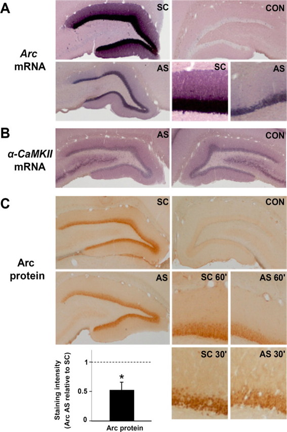
LTP reversal is coupled to rapid knockdown of upregulated Arc mRNA and protein expression. Arc AS- or SC-ODN was infused 2 h after LTP induction, and the brain was fixed by transcardial perfusion 1 h later. A, Coronal sections processed for in situ hybridization using a digoxigenin-labeled Arc riboprobe show robust upregulation of Arc mRNA in granule cell somata and dendrites in SC-treated controls. Arc mRNA expression was strongly inhibited after Arc AS treatment. The bottom right panels show high-magnification images of AS-treated and SC-treated dentate gyrus. CON, Contralateral nontreated dentate gyrus. These are representative images based on five experiments in each treatment group. Images were obtained from the mid-dorsal dentate gyrus within ∼300 μm of the recording site. B, α-CaMKII mRNA expression in Arc AS-treated and contralateral dentate gyrus. C, Arc immunohistochemical staining. Arc expression in dentate molecular and granule cell layer was reduced in AS-treated rats relative to time-matched, SC-treated control. High-magnification images of Arc staining at 60 and 30 min after AS or SC infusion at 2 h after HFS are shown in the bottom right panels. The bar graph shows the quantitative analysis of immunohistochemical staining in the dentate molecular layer at the 1 h time point. Optical density measurements were obtained along a line extending across the molecular layer of the inner blade of the dentate gyrus and normalized to background staining in stratum radiatum of CA1. Changes in Arc protein expression after AS treatment are normalized to SC-treated control. *p < 0.05.