
Figure 7.
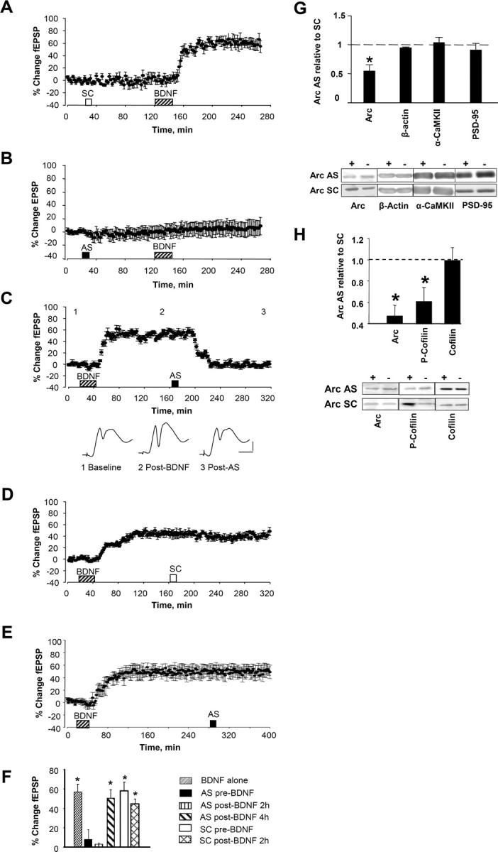
Arc AS blocked the induction and consolidation of BDNF-induced LTP. Time course plots show changes (means ± SEM) in the medial perforant path-evoked fEPSP slope expressed in percentage of baseline. Arc AS oligodeoxynucleotide or SC Arc sequence were infused 90 min before BDNF. A, Robust BDNF–LTP was induced in SC-treated rats (n = 5; p < 0.05). B, Arc AS pretreatment abolished BDNF–LTP (n = 6; p > 0.05). There was no significant difference in fEPSP slope values obtained immediately before and 2 h after BDNF infusion (p > 0.05). C, Top, Arc AS infusion at 2 h rapidly reverses ongoing BDNF–LTP (n = 5; p < 0.05). Bottom, Averaged field potential traces (4 sweeps) collected at the times indicated (1. baseline; 2, after BDNF; 3, after AS). Calibration: 2 mV, 2 ms. D, Infusion of SC Arc ODN at 2 h has no significant effect on ongoing BDNF–LTP (n = 5; p > 0.05). E, Arc AS infusion at 4 h had no significant effect on BDNF–LTP maintenance (n = 6; p > 0.05). F, Magnitude of fEPSP slope and population spike changes. n = 5–7 in all groups. *p < 0.05. G, Quantification of Western blots from dentate gyrus homogenates. Tissue was collected at the end of the experiments shown in A and B. Expression of Arc, but not β-actin, α-CaMKII, or PSD-95, were significantly reduced in AS-treated rats (*p < 0.05). Representative immunoblots below. + indicates infused dentate gyrus; − indicates contralateral, non-infused dentate gyrus. H, Dentate gyrus was microdissected at the end experiments shown in C and D, and homogenate samples were analyzed by quantitative Western blot. Mean ± SEM changes are expressed as AS-treated versus SC-treated dentate gyrus (*p < 0.05; n = 6–7 in all groups). Representative immunoblots are shown below. + indicates infused dentate gyrus; − indicates contralateral, non-infused dentate gyrus.