
Figure 16.
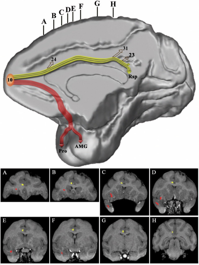
Three-dimensional reconstruction of the MRI of a macaque monkey brain to illustrate the course of the cingulate fasciculus (in yellow) that connects frontopolar area 10 with rostral and caudal cingulate cortex and the retrosplenial cortex and the uncinate fasciculus (in red) that connects area 10 with the rostral-most part of the superior temporal gyrus and the amygdala. The accompanying coronal sections (A–H) were taken at the levels indicated at the top of the medial hemisphere to show the course of these pathways as seen in coronal planes.