
Figure 2.
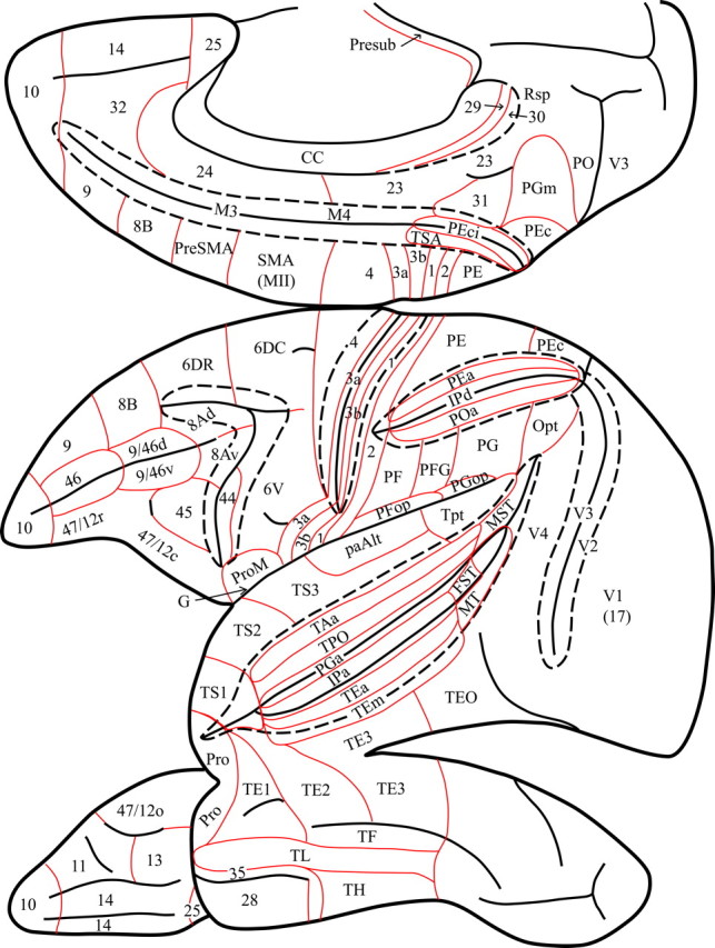
Diagram of the medial, lateral, and ventral views of the monkey left cerebral hemisphere to show the location of the architectonic areas used in the present article to describe the results. Interrupted lines indicate the lips of the opened sulci to show architectonic areas within them. Red lines show the borders of architectonic areas. Note the interrupted line that indicates removal of the posterior part of the corpus callosum to show the retrosplenial areas 29 and 30 that lie within the callosal sulcus.