
Figure 1.
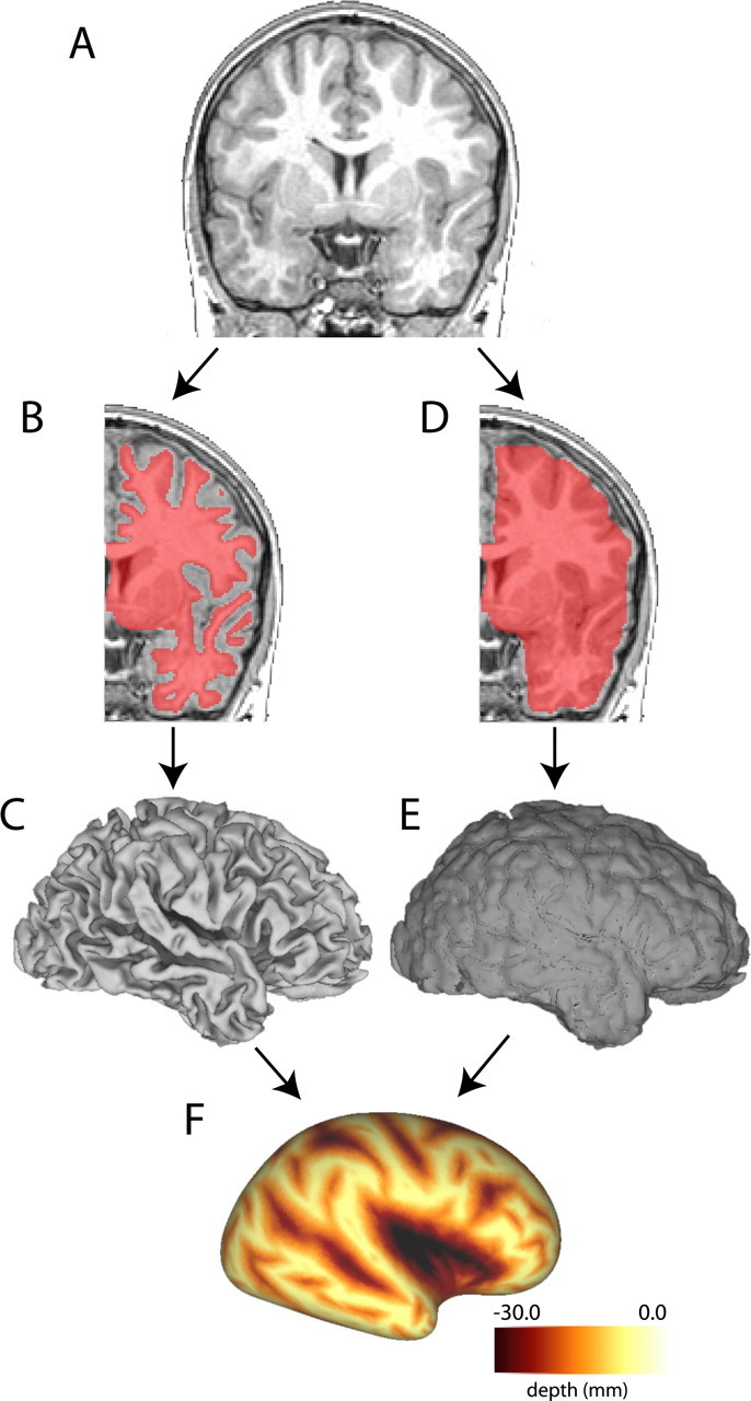
Method for generating sulcal depth maps. A, Coronal section through a high-resolution T1-weighted MRI for an individual subject. Hemispheres are cropped and analyzed separately. B, A segmentation boundary running midway through cortical gray matter (shown overlaid on the MRI in pink) is used to generate a fiducial surface (C). D, A cerebral hull boundary is created by dilating and expanding the segmentation boundary and a cerebral hull surface is generated (E). F, Sulcal depth maps are generated by calculating the distance from each node on the fiducial surface to the nearest node on the cerebral hull surface. Darker areas represent deeper sulcal depth measurements.