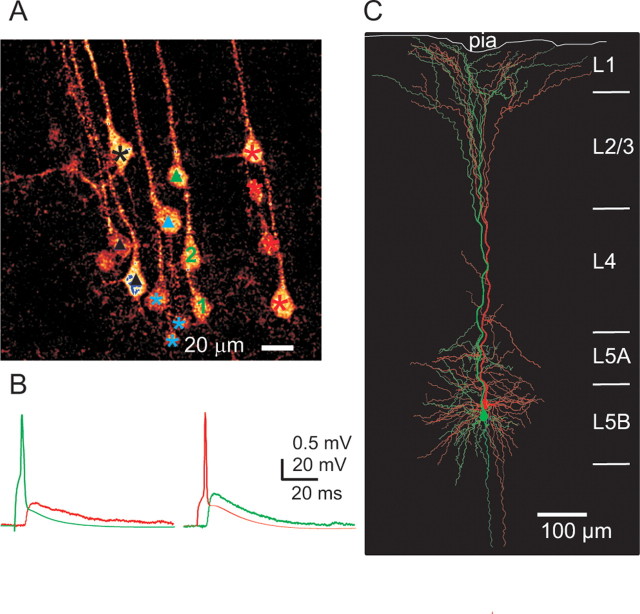
Figure 6.
Connected pair of layer 5B pyramidal cells located in the same cluster. A, Two-photon image from a living slice showing L5B pyramidal cells with apical dendrites projecting in bundles toward the pial surface. Cells marked by the same color symbol (black, blue, green, and red) identify cells belonging to the same cluster. Triangles and numbers mark cells recorded from. A reciprocal connection existed between cell 1 and cell 2. None of the other tested cells (triangles) were connected to cell 1 (connections to cell 2 not tested). B, Eliciting an action potential in cell 1 (green) evoked an EPSP in cell 2 (red) and vice versa. Calibration: EPSP trace, 0.5 mV, 20 ms; AP trace, 20 mV, 20 ms. C, Reconstruction of connected L5B pyramidal cell 1 (green) and cell 2 (red).

