
Figure 4.
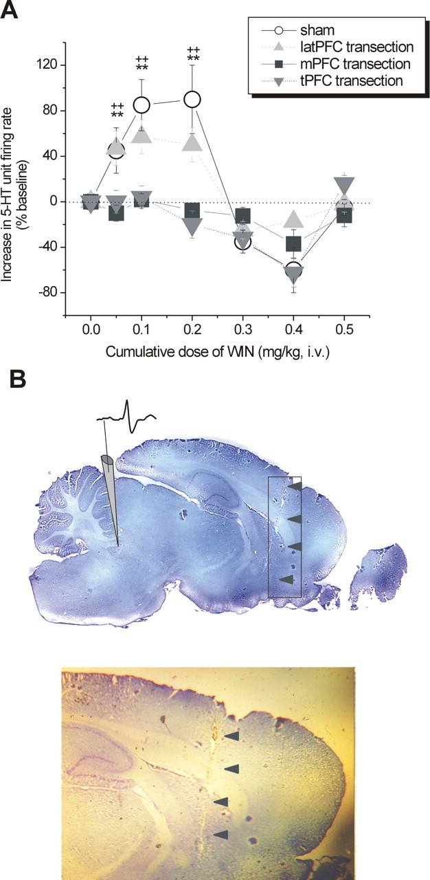
Effect of PFC transections on the modulation of DR 5-HT neuronal activity by intravenous administration of WIN. A, Line graph showing the modulatory effect of cumulative doses of WIN (0–0.5 mg/kg) on 5-HT single-unit firing activity after tPFC (shaded inverted triangles), ablation of mPFC (shaded squares), or latPFC (shaded upright triangles) subregions compared with sham-exposed controls (open circles). tPFC and mPFC transections abrogated the excitatory response to low doses of WIN (0.05–0.2 mg/kg, i.v.), whereas latPFC transection did not significantly reduce the excitatory response to low doses of WIN (0.05–0.2 mg/kg, i.v.). n = 3–7 animals per group. Values are expressed as mean ± SEM of increase in 5-HT unit firing rate (percentage of baseline). **p < 0.01 mPFC transection versus control; ++p < 0.01 tPFC transection versus control. B, Histological verification of the lesion left by a tPFC transection. Gray rectangle encompasses the anteroposterior range of all transections, and arrows point to an example of a cortical lesion trace on a midsagittal brain section (∼0.4 mm lateral to midline according to Paxinos and Watson, 1986). Shown is an illustrative depiction of the electrode placement and a typical action potential waveform of a putative 5-HT neuron (top) and a closer inspection of the lesion trace (bottom).