Figure 7.
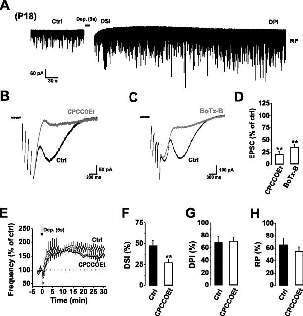
Autocrine mGluR1 activation occurs throughout early development. A, Continuous recording of mIPSCs from P18 PCs in control aCSF before and after a train of stimuli [Dep., +70 mV, 5 × 1000 ms (5 s stimulus) with 10 ms intervals] inducing DSI, DPI, and RP. The depolarization-induced inward current has been removed to improve clarity. B, C, Typical mGluR1-mediated slow EPSCs in response to a short train of depolarizing stimuli (5 × 50 ms stimuli at 10 Hz) in the absence (Ctrl) and presence of CPCCOEt (50 μm) (B) or after internally applying BoTx-B (500 nm) (C). For clarity, the stimulus artifacts have been removed. D, Bar graph of slow EPSC amplitudes in the presence of CPCCOEt (50 μm) or BoTx-B (500 nm). E, Normalized mean mIPSC frequencies before and after application of a 5 s stimulus in control aCSF (open circles) and in the presence of 50 μm CPCCOEt (filled circles). F–H, Bar graphs displaying the magnitude of DSI (F), DPI (G), and RP (H) after a 5 s stimulus in the absence (Ctrl) and presence of CPCCOEt (50 μm). **p < 0.01.

