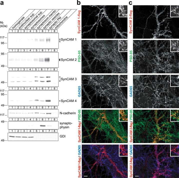
Figure 4.

SynCAMs are synaptic plasma membrane proteins present at excitatory but also found at inhibitory synaptic specializations. a, Synaptic plasma membrane fractionation of SynCAM proteins. The indicated subcellular fractions were prepared from rat forebrain at P9. Thirty micrograms of each fraction were analyzed by immunoblotting for the four SynCAM proteins as indicated. The synaptic membrane protein N-cadherin, the synaptic vesicle protein synaptophysin, and the soluble protein GDI were markers for these respective fractions. The numbers on the left indicate positions of molecular weight markers. Arrowheads show the running positions of the indicated SynCAM proteins in their predominantly adult (black) and postnatal (white) glycosylation forms. b, Exogenously expressed SynCAM 1 is sorted to both excitatory and inhibitory synaptic specializations in dissociated hippocampal neurons. Epitope-tagged SynCAM 1-flag was transfected into dissociated hippocampal neurons at 7 d.i.v. After transfection, mature cultures were analyzed at 21 d.i.v. by confocal microscopy after triple immunostaining for flag, the inhibitory presynaptic vesicle marker GAD65, and the postsynaptic excitatory marker PSD-95. Individual immunostainings are shown in the gray scale panels as indicated. The two panels at the bottom depict the indicated double merged images (SynCAM 1-flag, red; PSD-95, green; GAD65, blue). Insets in the top right of each panel represent 10-fold enlarged areas of the image shown. Confocal images were analyzed using Matlab for the number of single puncta and the occurrence of their double colocalization (SynCAM 1-flag, 2461 puncta analyzed; 3 images). Double colocalization identifies 53 ± 23% of SynCAM 1-flag puncta as immunopositive for PSD-95, and 24 ± 12% of SynCAM 1-flag puncta as immunopositive for GAD65. Errors are stated as SDs. The remaining SynCAM 1-flag puncta are removed from the synaptic markers analyzed here and nonsynaptic. We observed an occasional colocalization of GAD65 and PSD-95 in our hippocampal cultures, quantitated as an ∼10% mismatch of excitatory and inhibitory synaptic specializations similarly as described previously (Anderson et al., 2004). Scale bar, 10 μm. c, SynCAM 2 colocalizes with excitatory and inhibitory synapse markers in cultured hippocampal neurons. Epitope-tagged SynCAM 2-flag was expressed and analyzed as described in a (SynCAM 2-flag, 1951 puncta analyzed; 4 images). Double colocalization identifies 62 ± 28% of SynCAM 2-flag puncta as immunopositive for PSD-95, and 31 ± 12% of SynCAM 2-flag puncta as immunopositive for GAD65. Scale bar, 10 μm. The immunostainings shown in b and c are representative of the quantification results.
