Figure 9.

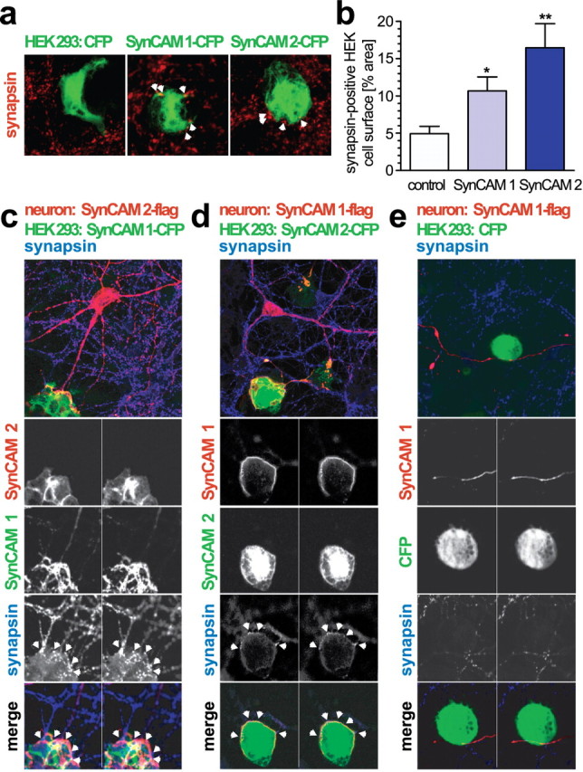
SynCAM 1 and 2 recruit presynaptic marker proteins and engage each other in adhesive neuronal complexes. a, SynCAM 1 and 2 recruit synapsin in a mixed coculture assay. HEK 293 cells expressing CFP-tagged SynCAM 1 or CFP-tagged SynCAM 2 were seeded atop dissociated hippocampal cultures at 9 d.i.v. HEK 293 cells expressing soluble CFP alone served as negative control. Cocultures were analyzed at 11 d.i.v. by confocal microscopy for localization of the presynaptic vesicle marker synapsin (red) and CFP (green). Merged stacks of serial optical sections are shown. Synapsin puncta were detected atop SynCAM 1- and SynCAM 2-expressing HEK 293 cells and are marked with white arrowheads in the images shown. b, Expression of SynCAM 1 or SynCAM 2 in HEK 293 cells cocultured with hippocampal neurons caused a significant increase of synapsin-positive puncta covering the cell surface area compared with control HEK 293 cells expressing CFP alone. The activity of both SynCAMs was similar (CFP alone, 4.9 ± 1.0% synapsin area coverage, n = 15 cells; SynCAM 1-CFP, 10.7 ± 1.9%, p = 0.011 relative to CFP, n = 15 cells; SynCAM 2-CFP, 16.5 ± 3.2%, p = 0.005 relative to CFP, n = 20 cells). Statistical analyses were performed using unpaired t tests with two-tailed p values. c, SynCAM 1 recruits and retains neuronal SynCAM 2 in adhesive neuronal complexes. Dissociated hippocampal neurons were transfected at 7 d.i.v. with flag-tagged SynCAM 2 to allow its visualization by immunostaining. HEK 293 cells expressing CFP-tagged SynCAM 1 were seeded atop these hippocampal cultures at 9 d.i.v., and cocultures were analyzed at 11 d.i.v. by confocal microscopy for localization of both SynCAM proteins and the presynaptic vesicle marker synapsin. Optical sections of each 1 μm thickness were obtained. The top panel shows, for a single optical section, the merged image (SynCAM 2-flag, red; SynCAM 1-CFP, green; synapsin, blue). The four panels at the bottom show for two serial optical sections the indicated individual immunostainings in grayscale and the triple merged image in color. White arrowheads mark synapsin puncta formed atop the SynCAM 1-expressing HEK 293 cell. d, SynCAM 2 reciprocally recruits neuronal SynCAM 1 into adhesive complexes. Analysis was performed as described in c in cocultures of hippocampal neurons expressing flag-tagged SynCAM 1 and HEK 293 cells expressing CFP-tagged SynCAM 2. The top panel shows, for a single optical section, the merged staining of the three indicated proteins (SynCAM 1-flag, red; SynCAM 2-CFP, green; synapsin, blue). The panels below show two serial optical sections obtained after immunostaining for the indicated proteins as described in c. e, Control HEK 293 cells do not retain SynCAM proteins. Analysis was performed as described in c in cocultures of hippocampal neurons expressing flag-tagged SynCAM 1 and HEK 293 cells expressing CFP alone. After surface contact of neurons expressing SynCAM 1, no synapsin retention or SynCAM complex formation was observed atop HEK 293 cells.
