
Figure 6.
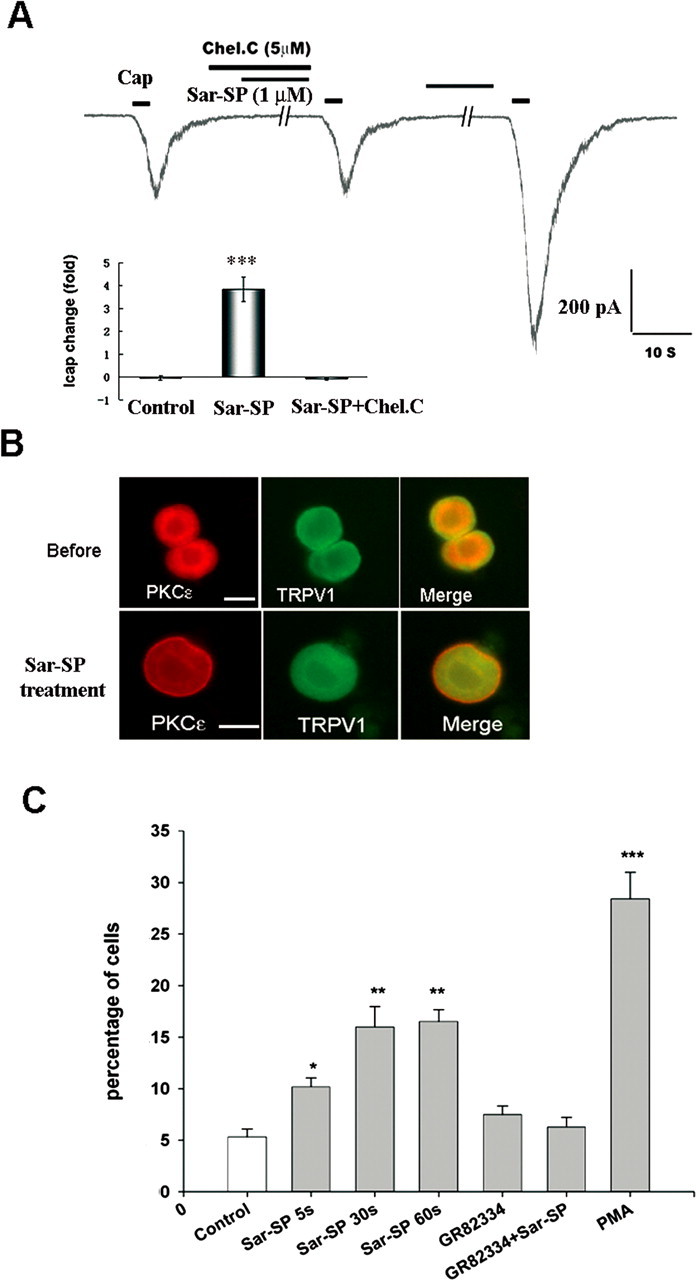
NK-1 activation induces membrane translocation of PKCε in DRG neurons that coexpress TRPV1. A, Involvement of PKC translocation in Sar-SP induced TRPV1 current enhancement. Coapplication of PKC translocation inhibitor Chel.C (5 μm) and Sar-SP (1 μm) failed to enhance the TRPV1 current. After washout, Sar-SP was still able to enhance the TRPV1 current. The inset summarizes the effect of Chel.C (***p < 0.001 vs control group; n = 15). Cap, Capsaicin. B, Double staining of PKCε (red) and TRPV1 (green) in dissociated DRG neurons before and after (1 min) Sar-SP (1 μm) treatment. PKCε and TRPV1 were heavily colocalized in small DRG neurons. Before Sar-SP (1 μm) treatment, PKCε was found in the cytoplasm, whereas TRPV1 was present both in the cytoplasm and on the membrane (top). After Sar-SP application, PKCε was greatly translocated to the plasma membrane. Scale bars, 20 μm. C, Percentage of neurons demonstrating PKCε membrane translocation after Sar-SP (1 μm for 5, 30, or 60 s). Pretreatment with the NK-1-specific antagonist GR82334 (1 μm, 15 min) completely prevented Sar-SP (1 min)-induced PKCε translocation. The PKC activator PMA serves as a positive control for the translocation (*p < 0.05, **p < 0.01, ***p < 0.001 vs the control group; n = 3).