
Figure 2.
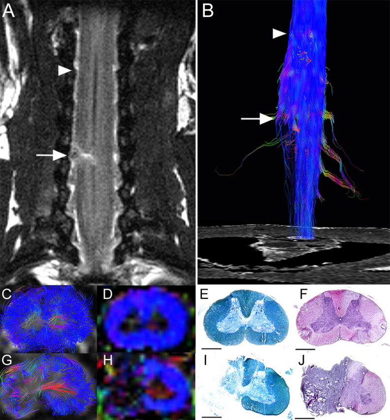
DTT of the hemisected spinal cord at 2 weeks after injury in a postmortem common marmoset. A, Coronal T2-weighted MRI depicted the hemisection injury as a low-intensity area with no change in the cord caudal to the injury. B, DTT of the hemisected spinal cord. The ROI was placed in the upper cervical spinal cord, and DTT was traced in the caudal direction revealing disruption of white matter fibers on the hemisected side. The traced tracts became untraceable at the injury site, whereas tracts on the contralateral side continued caudally. Arrows indicated the hemisection site and arrowheads indicated the point 8 mm cranial to the injury site in A and B. C–J, DTT (C, G), FA map (D, H), LFB staining (E, I), and HE staining (F, J) of the spinal cord 8 mm cranial to the injury site (C–F) and at the hemisection site (G–J). Although normal FA and anatomy of the spinal cord was confirmed cranial to the hemisection site, there was a significant decrease in FA of the white matter fibers at the hemisection site (G, H). Consistent with these changes in DTT (G) and color-coded FA map (H), demyelination was seen at the hemisection site (I). Scale bars, 1 mm.