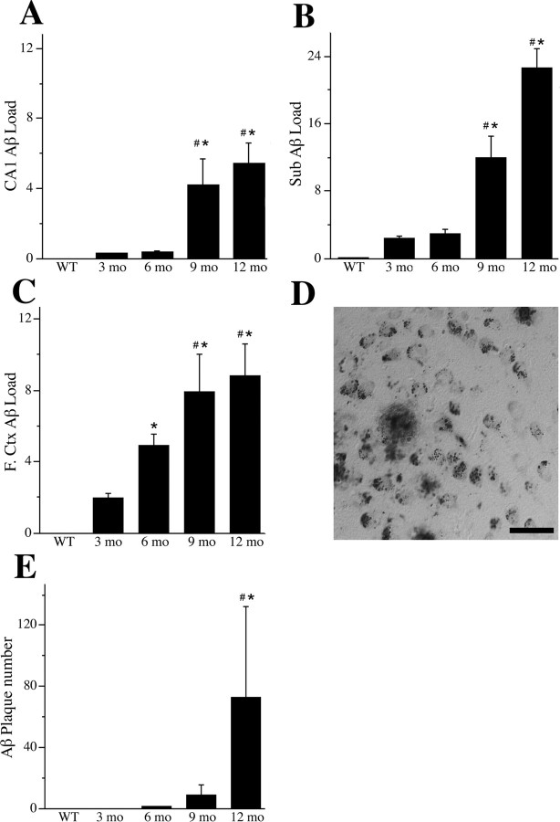
Figure 2.
Quantification of age-related increase in Aβ accumulation. Aβ load values were quantified in hippocampus CA1 (A), subiculum (Sub.; B), and layers IV and V of frontal cortex (F.Ctx.; C) from WT mice at 6 months of age and from female 3xTg-AD mice at 3, 6, 9, and 12 months of age. Data represent mean ± SEM Aβ load values from two to three nonoverlapping fields per section, 6–12 sections per brain (n = 7 per condition). ANOVA showed significant between group differences in Aβ load in hippocampus CA1 (F = 5.98, p = 0.008), subiculum (F = 31.85, p < 0.001), and frontal cortex (F = 7.6, p = 0.008). D, Aβ-IR also included extracellular Aβ deposits that were observed primarily in subiculum. Scale bar, 50 μm. E, Data show mean ± SEM counts of extracellular Aβ deposits in subiculum and hippocampus CA1 from 12 sections per animal at ages 3, 6, 9, and 12 months in 3xTg-AD mice and age 6 months in WT mice. A significant age-related increase in Aβ deposits was observed in 3xTg-AD mice (F = 9.70, p < 0.001). *p < 0.05 compared with 3 months; # p < 0.05 compared with 6 months.

