Figure 2.
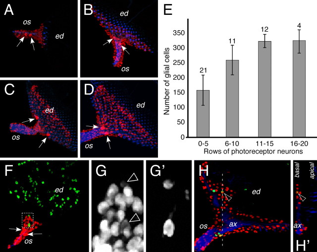
Formation of the eye disc glia. A–D, F–H, Larval eye imaginal disc stained for the presence of glial nuclei (Repo staining, red), neuronal membranes (HRP staining, blue) and dividing cells (phospho histone H3 staining, green). A–D, Glial cells are born in the optic stalk and migrate onto the eye imaginal disc as neuronal differentiation proceeds. Two large glial nuclei can be recognized already from early developmental stages onwards (arrows). Initially these nuclei are found in the optic stalk (os), but later are generally located at the posterior end of the eye imaginal disc (ed). Note that glial nuclei are always posterior to the differentiating neurons. E, Glial nuclei were counted using the Volocity classifier tool. The number of eye discs quantified is indicated. Discs were staged according to the number of rows of photoreceptor cells. At the end of the third instar larval stage, ∼325 glial cells can be detected in the eye imaginal disc. F, Eye imaginal disc of a second instar larvae. No neuronal differentiation can be detected in the eye imaginal disc. Dividing cells are detected after a phosphohistone H3 staining. Some glial cells divide in the stalk and generally show reduced levels of Repo expression (dashed area is shown in high magnification in (G). G, Repo expression highlights all glial nuclei. Arrowheads indicate dividing glial cells that (G′) are labeled after a phosphohistone H3 staining. H, In third instar eye imaginal discs, glial cells divide in the optic stalk (os). In addition basal glial cells located in the disc can also divide (arrowhead). H′ shows an orthogonal section at the position indicated by the white dashed line in (H. ax, Axons.

