Figure 4.

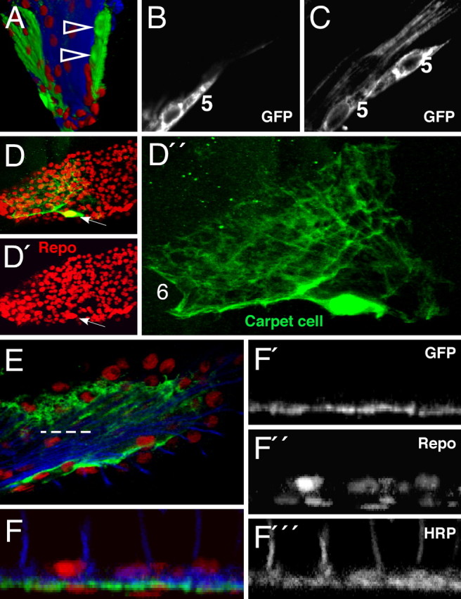
Marginal and carpet glial cells. Confocal analysis of third instar larval eye imaginal discs. Single glial cells are marked with anti-GFP antibodies (green). Glia cell nuclei are stained for Repo expression (red) and photoreceptor neurons are marked with anti-HRP antibodies (blue). A–E, Anterior is to the top. A, 3D reconstruction of an eye imaginal disc. At the margin of the eye disc, a small clone of glial cells is marked (arrowheads). B, C, Single confocal sections of this glial clone show that marginal glial cells (5) display an elongated, clapboard like shape. No contacts to axons can be detected. D, D′, Every eye disc has two large, flat carpet cells of which one is labeled in this eye disc. The carpet glial nucleus can be easily identified according to its size (arrow; see also arrows in Fig. 2). The cytoplasmic extensions of the carpet cells cover half of the eye disc. E, Triple-stained specimen showing the relation of the carpet cell to axons and other glial cells. The dashed line indicates the orthogonal section shown in F. F–F‴, Orthogonal section shows that the so-called carpet cells separate basally localized nuclei from axons (blue) and more apical located glial nuclei.
