
Figure 6.
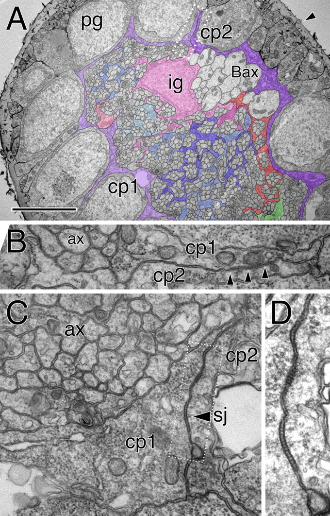
Glial cells of the optic stalk. Electron microscopic images taken from an optic stalk of a third instar wandering larvae. A, The optic stalk is surrounded by a thick neural lamella (arrowhead). The perineurial glial cells (pg) have a cuboid shape with some fine cell protrusions that extend along the axis of the stalk. Below the perineurial cells are two subperineurial cells (cp1, cp2) that are highlighted by colored shading. These cells form a continuous thin ring around the entire optic stalk. The axonal profiles of the Bolwig's organ can be recognized because of their size (Bax). One inner glial cell nucleus is seen (ig). Some glial processes that enwrap individual axons are highlighted. Here, glial processes do not surround axons at the periphery of the stalk. The boxed area is shown in higher magnification in B. Scale bar, 3.5 μm. B, High magnification of the carpet glia contact zone. Septate junctions can be recognized (arrowheads) between carpet cell one and two (cp1, cp2). Axonal profiles (ax) have not yet been engulfed by glial processes. C, D, Similar images of the septate junction area between two carpet cells (arrowhead, sj). The corresponding optic stalk is shown in supplemental Figure 3 (available at www.jneurosci.org as supplemental material).