
Figure 1.
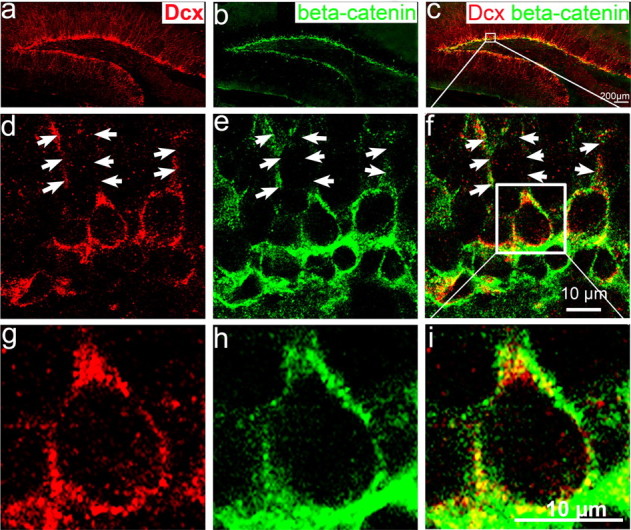
β-Catenin is expressed in postnatal dentate gyrus and colocalizes with Dcx-positive newborn neurons at the inner granular cell layer. Immunohistochemistry of hippocampal sections from wild-type mouse brain at P25 labeled with antibodies to the newborn neuronal marker Dcx (red) and to β-catenin (green). a, Dcx positive-cells located in the inner cell layer of dentate gyrus. b, β-Catenin is highly expressed in the SGZ and inner granular cell layer. c, Merged image of a and b showing that β-catenin is expressed in Dcx-positive newborn neurons adjacent to the SGZ. d, e, Confocal images of single focal section further shows that Dcx (d) and β-catenin (e) are highly expressed in the cytoplasm and in the processes of newborn neurons (arrows). f, Merged image of e and d showing that β-catenin colocalizes with the Dcx-positive newborn neurons in the dentate gyrus. g–i, Enlarged view of single newborn neuron within the white box in f showed that Dcx-positive newborn neurons (g) expresses β-catenin (h) in the cytoplasm (i), but not detectable in the nucleus.