Figure 1.
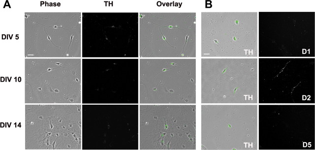
Characterization of DA neurons in VTA cultures prepared from P1 rats. A, DA neurons were identified based on TH staining. After 5, 10, or 14 DIV, cultures were permeabilized and incubated with TH antibody followed by secondary antibody conjugated to Alexa 488. B, DA neurons in VTA cultures express D2 receptors, but not D1 or D5 receptors on the cell surface. Cell surface DA receptors were labeled by incubating live cells (10 DIV) with polyclonal antibodies recognizing the extracellular N-terminal of the D1, D2, or D5 receptor, followed by secondary antibody conjugated to Cy3. Then, cells were permeabilized and stained for TH followed by secondary antibody conjugated to Alexa 488. Left, Green indicates TH+ neurons; right, cell surface DA receptor staining. Scale bars: 20 μm.

