Figure 2.
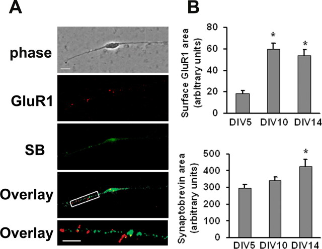
DA neurons in VTA cultures express cell surface GluR1 mainly at extrasynaptic sites. Cell surface GluR1 was labeled by incubating live cultures with polyclonal antibody recognizing the extracellular N-terminal domain of GluR1, followed by Cy3 conjugated secondary antibody. Then cultures were permeabilized and immunostained for the synaptic marker synaptobrevin/VAMP 2, followed by Alexa 488 conjugated secondary antibody. A, Phase image, GluR1 surface expression (red), synaptobrevin staining (SB, green), overlay of GluR1, and synaptobrevin staining (yellow) and a higher-magnification overlay of a 30 μm section of a neuronal process. Scale bar: top four panels, 10 μm; bottom, 5 μm. B, Qualification of surface GluR1 staining area (top) and synaptobrevin staining area (bottom) for DA neurons on different DIV (n = 19–30; Dunn's test, *p < 0.05 compared with DIV 5). Error bars indicate SEM in this and all subsequent figures.

