Figure 8.
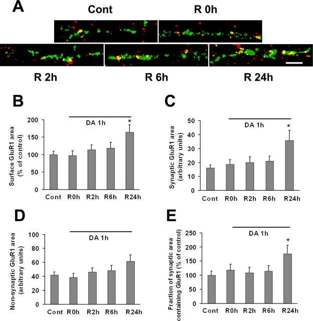
GluR1 surface and synaptic expression by DA neurons is increased 24 h after prolonged (1 h) incubation with DA. VTA-PFC cocultures were treated with 1 μm DA or media for 1 h and then allowed to recover in media for 0 h (R0 h), 2 h (R2 h), 6 h (R6 h), or 24 h (R24 h). A, Representative images show overlay of surface GluR1 (red) and synaptobrevin (green) staining after different recovery times. Scale bar, 5 μm. B–E, Quantification of surface GluR1 staining (B), synaptic GluR1 area (C), nonsynaptic GluR1 area (D), and the portion of synaptic area containing GluR1 (E). After 24 h recovery, all measures except nonsynaptic GluR1 area were significantly increased (n = 20–23; Dunn's test, *p < 0.05).

