Figure 9.
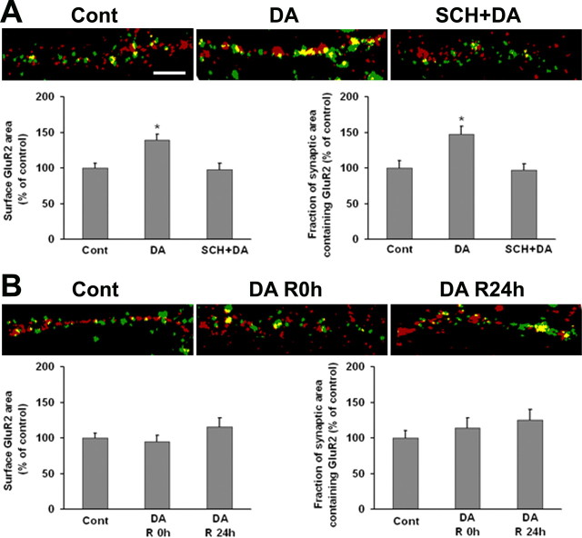
GluR2 surface and synaptic expression by DA neurons is increased after brief (10 min), but not prolonged (1 h) incubation with DA. A, VTA-PFC cocultures were treated with media, DA (1 μm, 10 min), or SCH+DA (10 μm SCH 23390 was added 5 min before DA). Representative images show overlay of surface GluR2 (green) and synaptophysin (red) staining. Bottom shows the quantification of surface GluR2 staining and the portion of synaptic area containing GluR2. DA increased surface and synaptic GluR2, this was blocked by SCH 23390 (n = 20–25; Dunn's test, *p < 0.05). DA also increased nonsynaptic GluR2; this was also blocked by SCH 23390 (data not shown). B, VTA-PFC cocultures were treated with 1 μm DA or media for 1 h and then allowed to recover in media for 0 h (R0 h) or 24 h (R24 h). Representative images show overlay of surface GluR2 (green) and synaptophysin (red) staining. The bottom shows quantification of surface GluR2 staining and the portion of synaptic area containing GluR2. In contrast to results obtained for GluR1 (Fig. 8), surface and synaptic expression of GluR2 were not altered after prolonged DA treatment (n = 20–24; ANOVA, p > 0.05). Nonsynaptic GluR2 was also unchanged (data not shown). Scale bar, 5 μm.

