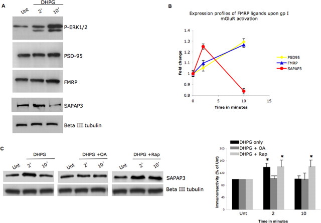
Figure 5.
Group I mGluR activity-dependent FMRP phosphorylation correlates with translation of FMRP ligand SAPAP3. A, Neurons were treated with 100 μm DHPG for 0 [untreated (Unt.)], 2, or 10 min, and protein expression levels of the FMRP ligands were examined by Western blotting cytoplasmic cell lysates. A synapse associated protein, SAPAP3, shows changes in protein expression that correlate with the time course of FMRP phosphorylation. B, Densitometry analyses of the changes in FMRP ligand expression in the presence of 100 μm DHPG in primary hippocampal neurons. Error bars represent the SD between sample triplicates in the assay. C, Neurons were treated with 100 μm DHPG for 0 (untreated), 2, or 10 min in the presence of DHPG alone/DHPG stimulation in the presence of 10 nm OA or 20 μm rapamycin. SAPAP3 protein expression levels as measured by Western blotting of cytoplasmic cell lysates show PP2A activity-sensitive changes in protein expression. In the right panel, SAPAP3 immunoreactivity was normalized to β-III tubulin under the conditions studied, calculated as ± SEM, and represented as a histogram (n = 4; the asterisk denotes significance compared with untreated with a Student's t test, *p < 0.05).

