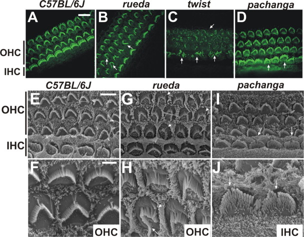
Figure 4.
Analysis of hair cell morphology in rueda, twist, and pachanga mice. A–D, Cochlear whole mounts of P5 animals were stained with phalloidin (green). Outer hair cells (OHC) and inner hair cells (IHC) are indicated. Hair bundle morphology and polarity were altered in rueda, twist, and pachanga mice (arrows). E–J, Cochlear whole mounts were analyzed by SEM. Kinocilia in hair bundles of rueda mice were frequently in ectopic positions (G, H, arrowheads), and hair bundles were asymmetric (H, arrow). Hair bundles in pachanga mice were often split (I, J, arrows). Scale bars: (in A) A–D, 8 μm; (in E) E, G, I, 5 μm; (in F) F, H, J, 2 μm.

